| Biography | Research | Publications | Teaching | Group | Projects | Software | Sponsors | Collaborators |

|

|
|
|
|
|
| (a) | (b) | (c) |

|
 trimer subunit 1 trimer subunit 1 trimer subunit 2 trimer subunit 2 trimer subunit 3 trimer subunit 3
|
|
|
|
|
| (d) | (e) | (f) |
|
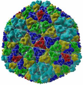
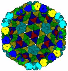
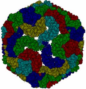
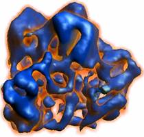
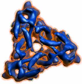
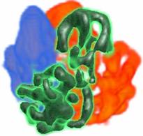
Figure (a), (b) and (c) are three visualizations of the same spherical nucleo-capsid half-shell model of the Rice Dwarf Virus (RDV), using combined surface and volume rendering. The different colors show the nucleo-capsid shell from the outside (a) and (c), and the inside (b), helps vividly bring out the local and global complexity of the quasi-symmetric packing of the individual structure protein units. Each individual structure unit, exhibiting a trimeric fold, is further visualized in (d), and (e) from two different views, as well as with different colors (f) showing the three different conformations of the monomeric protein chain forming the trimeric structure unit. Additional images for Virus-modeling Additional movies for Volume Rover demonstration |
||